Degenerative intervertebral discs (technical)
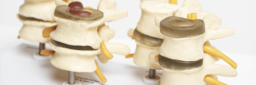
Degenerative intervertebral discs describe the changes associated with a loss of structural integrity within an intervertebral disc. These changes may occur during the normal ageing process or accelerated with lifestyle, sporting, nutritional, occupation and hereditary factors. Pathological changes within discs that lead to accelerated degenerative changes include inflammatory diseases, infection and trauma.
Three things that must be seen to classify a disc as degenerative and include:
- Intervertebral disc annular fissures
- Intervertebral disc degeneration
- Intervertebral disc herniation
1. Intervertebral disc annular fissures
Annular fissures are sometimes called annular tears. The term ‘tear’ is not appropriate concerning degenerative discs. The word tear implies trauma. An annular fissure is a term used to describe either a separation between the fibres of the outer disc (annulus fibrosis – annulus for short) or a separation between the annulus and the vertebral bones (ring apophysis). Annular fissures can be described by their orientation. These include:
- Concentric or circumferential. Fissures are between the layers of the annulus.
- Radial fissures. Fissures extend through the annulus. These fissures can run horizontally, obliquely or vertically.
- Transverse fissures. Transverse fissures separate the disc (annulus) from the bone (apophyseal bone)
If these fissures large and separation of annular fibres is gross, fissures can be called annular gaps.
Diagnosing intervertebral disc fissures
MRI is the imaging modality used to diagnose fissures. On MRI any dehydration (looks black on MRI) suggests at least one fissure. There may be several. The bright white appearance of a disc on MRI (high-intensity zones – HIZ) suggests fluid or granulation tissue (young repair tissue) associated with a fissure. This white appearance can be highlighted by injecting gadolinium into a disc and taking another MRI. Not all fissures are observed on MRI.
- Painful fissures may not be seen on either MRI or CT scan.
- Not all tears observed on imaging are symptomatic. Non-painful discs also contain tears observable on MRI and CT discogram. Therefore to assume all tears in discs are painful is incorrect.
2. Intervertebral disc degeneration
Intervertebral disc degeneration implies wear and tear or break down of a disc. It can be natural or pathological. As disc ages and becomes degenerative, one or more of the following will be seen.
- Intervertebral disc desiccation (dehydrated – Black on MRI)
- Intervertebral disc fibrosis (scar tissue within it)
- Intervertebral disc space narrowing (disc becomes thin and loses its suppleness)
- Intervertebral disc bulging (Bulges out from losing its ability to support weight)
- Intervertebral fissuring (tears within a disc)
- Mucinous degeneration of the annulus (change in the material of the disc – now easier to tear)
- Intervertebral disc intradiscal gas (gas accumulates in the gaps that tears create in the fibres of the annulus)
- Osteophytes of the vertebral apophyses (inflammation leads to bone spurs – osteophytes)
- Sclerosis of the end plates (The vertebral bones above and below the disc become stiffened and hardened)
3. Intervertebral disc herniation
The term disc herniation means there has been some displacement of intervertebral disc material beyond the intervertebral disc space. The intervertebral disc space is bordered by the vertebra (endplates above and below) and around its circumference the outer edge of the ring apophyses. Disc material includes the nucleus, cartilage, apophyseal bone and annular tissue. The displaced content may be one of these structures or a combination. Disc herniation is not a synonym for an exclusive nucleus pulposus herniation as it may contain other structures.
Disc bulging
Disc bulging is not a synonym for a disc herniation. Disc bulging is the term used to describe the extension of the whole circumference of the disc extending beyond to ring apophyses (generally more than 25% of disc fibres) and can be asymmetrical.
Herniated discs
There are two types of herniated discs and are classified on the basis of their shape (of displaced material).
- Disc protrusion
A disc protrusion involves less than 25% of the disc circumference. The term for this is ‘focal’. A disc protrusion is wider at the base (origin, at the start of the protrusion within the disc) than the width of the protruded material extending from the disc. The protruded intradiscal material must be continuous with the protruded disc material. That is, there is no separation.
- Disc extrusion
In a disc extrusion, the width of the herniated material is larger outside the disc space than inside the disc. A subcategory of disc extrusion is disc sequestration. Sequestration occurs when there is no longer continuity between the intradiscal substance and the extruded disc substance.
It is worth highlighting. Disc herniations may also occur in a vertical direction. Vertical herniations will go through a gap in the vertical body endplate. It is sometimes called a Schmorl node.
You may also be interested in reading these articles by our Balmain and Rozelle chiropractors
- Intervertebral disc summary
- Degenerative intervertebral discs (non-technical)
- Normal Vs pathological intervertebral disc changes
- Intervertebral disc bulge
- Intervertebral disc herniation
- Intervertebral disc protrusion
- Intervertebral disc extrusion
- Intervertebral disc sequestration
- Intervertebral disc fissures
- Intervertebral disc tears
- Intervertebral disc infection
- Traumatic intervertebral disc injury


